What is IRM microscopy?
Interference reflection microscopy (IRM) is an optical microscopy technique that takes advantage of polarized light to form an image of an object on a glass surface1-2. The intensity of the signal is a measure of the proximity of the object to the glass surface. This technique can be used to study cells or microtubules at the surface without the use of fluorescent labels.
It can be difficult to apply regular reflection microscopy to study cells or microtubules. Relatively high-power laser light is used to form an evanescent wave with sufficient energy. This results in scatter reflections, and refractions between the media producing undesirable rays of light that are called “stray light”. Stray light contaminates the exponential decay of the evanescent wave, excites the bulk of the specimen, and deteriorates the TIRF effect.
IRM uses a phenomenon known as wave interference, and more specifically, the interference of polarized light. As a result of the interference, the cell–glass interface appears as various shades of gray with increasing darkness, which indicates the relative proximity between a cell and the coverslip. The darkest areas are the result of destructive interference at the cell‒glass interface, and the lighter areas result from different degrees of constructive interference. IRM is the easiest and fastest method to obtain immediate information about the overall focal adhesion distribution, which represents the cell fingerprint at the coverslip level. Since the method does not employ fluorescence labeling, cell–substrate separation distances can be determined even in unstained cells
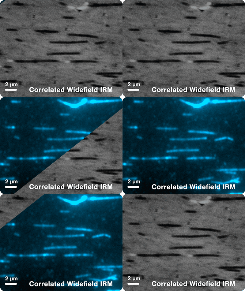
The figure shows fluorescently-labeled microtubules imaged using IRM and Widefield. Imaged with an integration time of 20 ms and 100 ms for IRM and Widefield, respectively.
How does it work in practice?
IRM works by using polarized light to form an image on the glass surface where the intensity of the signal measured corresponds directly to the proximity of the object to the glass surface. Light of a specific wavelength is first passed through a linearly polarizing filter, the light is then reflected by a beam splitter towards the objective which focuses it on a sample over the glass. The glass surface will reflect the polarized light but allow unpolarized light to enter and reach the sample and be reflected off of it. Depending on the experimental configurations, the light can then either be:
- Reflected back being phase-shifted half a wavelength, and therefore destructively interfere with incoming light which is observable as dark spots in the final image.
- Reflected back with a similar wavelength and result in brighter regions in the final image.
- Not contact the sample and continue after reflecting from the glass, appearing as bright pixels.
The reflected light will travel back to the beam splitter and pass through a second polarizer, which eliminates scattered light, before reaching the detector in order to form the final picture.
Explore other imaging techniques
Widefield
Image biomolecular processes with low protein concentration in solution with high acquisition rates.
Confocal Fluorescence
Multicolor confocal, perfect for visualizing biological processes in solution.
STED Super Resolution
The perfect choice for performing experiments in highly crowded environments, offering unprecedented resolution (< 35 nm).
TIRF Fluorescence
Suitable for visualization on surfaces as it eliminates background fluorescence outside the focal plane.
References
1. Simmert et al. (2018) LED-based interference-reflection microscopy combined with optical tweezers for quantitative three-dimensional microtubule imaging. Optics Express
2. Mahamdeh et al. (2018) Label-free high-speed wide-field imaging of single microtubules using interference reflection microscopy. Journal of Microscopy
Our solution
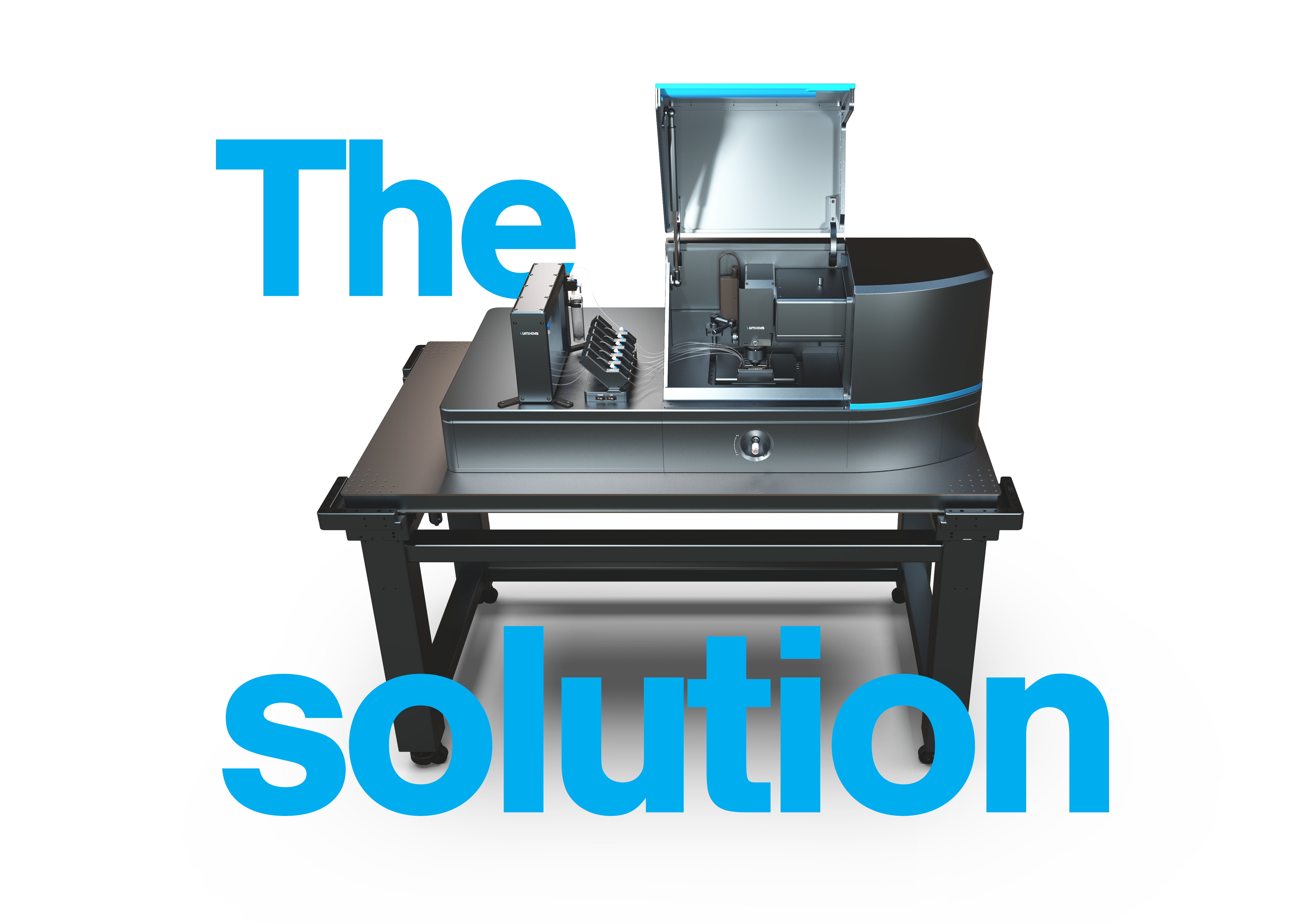
The C-Trap® Optical Tweezers – Fluorescence & Label-free Microscopy is the world’s first instrument that allows simultaneous manipulation and visualization of single-molecule interactions in real time. It combines high resolution optical tweezers, fluorescence and label-free microscopy and an advanced microfluidics system in a truly integrated and correlated solution.
The C-Trap offers you a fast workflow to seamlessly catch and manipulate single molecules. The instrument measures their structural changes or interactions while you visualize them in teal time with high spatial and temporal resolution, ultimately offering you a complete and detailed picture of biomolecular properties and interactions
Curious to learn more?
