Study protein folding and conformational dynamics at the nanoscale

Part of: Protein folding

Study protein folding and conformational dynamics at the nanoscale

C-Trap Protein Folding

Study of multi-domain protein folding & conformational dynamics

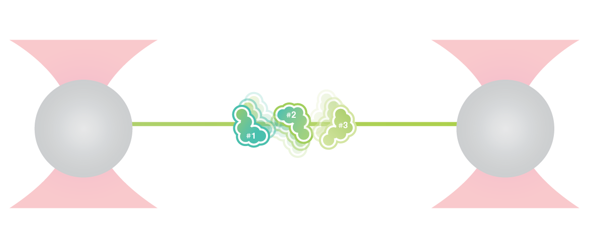
In this experiment, a protein is tethered between two optically trapped beads. The folding and unfolding of the protein is controlled by moving the beads while the force and distance are measured simultaneously.

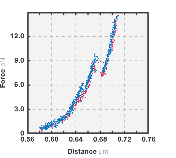
The resulting force-distance curve (Figure 1) of calmodulin – a calcium-binding protein – reveals that the protein unfolds and refolds in two steps which correspond to two separate helix-loop-helix domains. By simultaneously observing the FRET fluorescence signal, it can be seen how the unfolding of the domains results in fluctuations of the FRET signal due to changes in the relative position of the FRET pair.

Equilibrium dynamics showing the transition between short-lived intermediate states can be studied because of the intrinsic distance clamp which keeps the traps at a fixed distance, while force fluctuations are measured. When this is applied to calmodulin, the equilibrium fluctuations between the states can be observed with a force resolution of <0.1 pN at 100 Hz. Figure 2 shows that calmodulin switches between two major states, without a clear preference and that intermediate steps can be resolved as calmodulin occasionally jumps to a third state for short periods of time, represented by the dashed grey lines.

1 Force-extension (blue) and force retraction (red) cycle of full length CaM at 10 mM Ca2+. Pulling and retraction speed is 100 nm/s. Bead diameter is 1.0 µm and trap stiffness is 0.284 pN/nm. The figure shows 200 Hz data.

2 Full length CaM protein at 10 mM Ca2+ showing equilibrium dynamics between multiple states. Data is recorded at 50 kHz (grey line) and averaged at 200 Hz (red line). The histogram quantifies the most populated states in the inset (right panel) showing two peaks at 6.5 ± 0.1 pN and 7.8 ± 0.09 pN (mean ± standard deviation).

Sample courtesy of Prof. Carlos Bustamante at the University of California, Berkeley.
Read more:
Mashaghi et al. (2016) Nature
Neupane et al. (2016) Science
Pelz et al. (2016) Nature Communications

Solutions

C-Trap® Optical Tweezers Fluorescence & Label-free Microscopy
C-Trap

Optical Tweezers and Fluorescence Microscopy

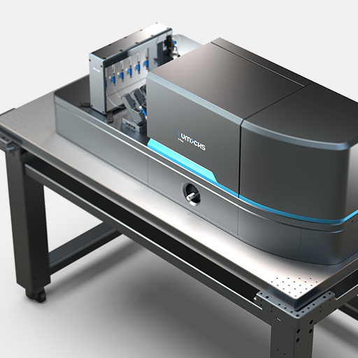
The C-Trap is the world’s first instrument that allows simultaneous manipulation and visualization of molecular interactions in real-time. It combines high-resolution optical tweezers, confocal microscopy, or STED nanoscopy with an advanced microfluidics system in a truly integrated and correlated solution.

M-Trap® Optical Tweezers Fluorescence & Label-free Microscopy
m-Trap

Optical Tweezers

The m-Trap is the first entry-level optical tweezers instrument specifically developed for high-resolution single-molecule research. Ultra-high force resolution and stability, with incredible throughput, ease of use and modularity ‒ all at an unprecedented price level.

Download the brochure to learn more

Join our mailing list

I would like to receive exclusive news about the latest product developments, dynamic single-molecule and immuno-oncology events and breakthrough science. I can revoke my consent at any time.

By submitting the form you agree to LUMICKS' privacy policy.

Close Menu

For pricing, reach out to your application scientist or account manager

Open Email

Fill in our contact form, we will reach out to you!

Please include ‘Lakeview data analysis price inquiry’ in the Message box

Contact Us


Join our newsletter

Get exclusive news on the latest publications, product developments, events and breakthrough science.

By submitting the form you agree to LUMICKS' privacy policy. You can revoke your consent at any time.

Download our webinar recording:


Download our Cell Therapy (CAR-T, TCR, NK) applications deck