Receptor responses to mechanical stimuli
In this experiment, we used the C-Trap to understand the properties involved in the mechanical activation of transmembrane receptors. While applying a controlled mechanical stimulus, we monitored the activation of intracellular responses and signals in real time.

We guided a microscopic bead to the cell surface of a human HEK293 cell expressing fluorescently labeled biotinylated receptors (blue signal) and cytosolic marker (red signal).
Figure 1 shows how moving the bead away from the cell surface with forces above 300 piconewtons caused deformation of the cell membrane and accumulation of receptors at the site of manipulation. As we proceeded, minimal applied forces on the cell membrane were enough to pull out a thick membrane tube.
These observations suggest a correlation between cell membrane deformation and local accumulation of receptor protein at the site of manipulation
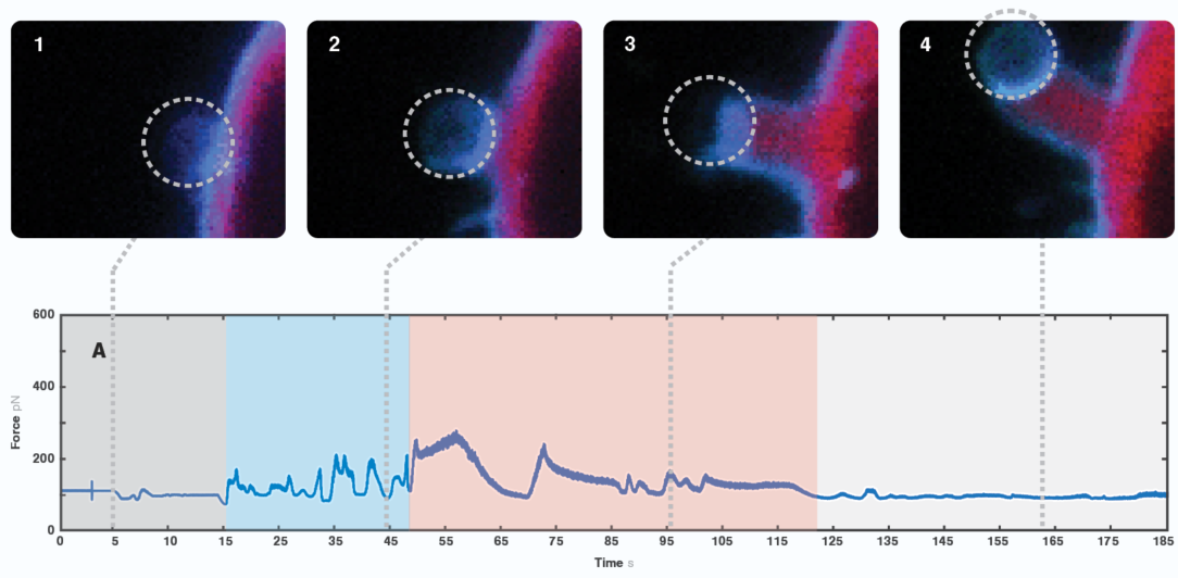
1 Confocal microscopy images showing cellular changes upon manipulation of the bead. Bottom graph shows measurements of forces exerted on the cell membrane over time.
Filopodia formation and functions
The following experiment highlights the forces and kinetics associated with filopodia formation. To assess the process, we studied Dictyostelium discoideum cells, a eukaryotic amoeba cell and model system that is commonly used to understand human cell processes.

We moved the bead in proximity to the D. discoideum cell, ectopically expressing GFP-Myosin 7 (blue signal) and actin filament marker RFP-LifeAct. The cell moved in the direction of the trapped bead, extending multiple protrusions towards it upon sensing the proximity of the object.
We observed a characteristic signal every time the cell engulfed the bead (Figure 2). Interestingly, we also found an occasional force spike right before bead engulfment. Since we observed the formation and retraction of one of the formed filopodia touching the bead, we suggest that this peak possibly is caused by the interaction between the cell protrusion and the bead. Thus, the maximum value shown on the graph (around 20 pN) corresponds to the force that a single protrusion exerted over the foreign object.
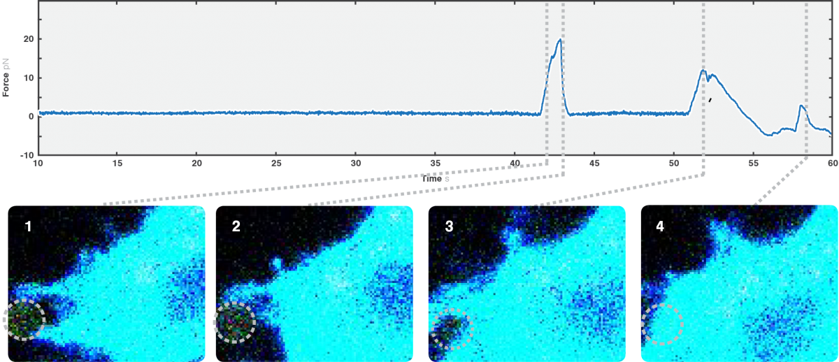
2 Confocal images of a cell interacting with a trapped bead, including GFP-Myosin 7 (blue signal) and actin filament marker RFP-LifeAct (green signal). The graph depicts the measured forces exerted by cell protrusions on the bead over time.
Cellular droplet fusion and small components in a multicellular organism
The C-Trap can also manipulate small components inside of living multicellular organisms. We used the optical tweezers to manipulate intrinsic lipid droplets and study droplet fusion inside of Caenorhabditis elegans (roundworm).
Using brightfield microscopy, we can quickly scan cell samples with a micrometer-precision stage and, on top of that, obtain fine movements when needed using a nanometer-precision positioning system. These features allow us to optically trap droplets in two ways: dragging the droplets around the body, thus exposing them to each other, or keeping them at the same position and letting the stage bring other droplets into the proximity of the trapped molecule.
The approach allowed us to follow the small components in vivo and in real time and assess forces associated with the fusion process (Figure 3).
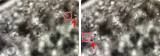
3 Brightfield microscopy imaging showing directional manipulation of a lipid droplet (indicated by the red arrow) with a laser (focused in the center of the red circle) inside the C. elegans.